Skip Navigation

Prenatal Form and Function – The Making of an Earth Suit

  
[Help]

Unit 5:   4 to 5 Weeks

  Closer Look: 
  Applying the Science: 
Play Movie - 4 to 5 Weeks
Movie 5.1 - 4 to 5 Weeks
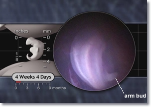
4 week 4 days embryo, arm bud
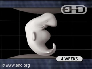
Figure 5.1 - Embryo From the Back
The Biology of Prenatal Development DVD
Copyright © 2006 EHD, Inc. All rights reserved.


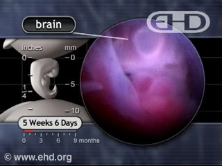
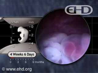
The brain continues growing at an incredible rate. Between 4 and 5 weeks, the 3 primary vesicles divide into 5 secondary vesicles.1 During this time, the head makes up about one-third of the embryo’s entire size.2 An early form of the cerebellum appears by 4 to 4½ weeks; this area of the brain will later control muscle control and coordination.
Play Movie - Cerebral Hemispheres
Movie 5.2 - Cerebral Hemispheres
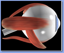
optic nerves, eye muscles
Figure 5.2 - Optic Nerve and Muscles of the Eye
The optic nerves connect your eyes to your brain, allowing you to see.
LifeART
Copyright © 2002 Lippincott, Williams & Wilkins.

By 4½ weeks portions of the brain forming the right and left cerebral hemispheres appear.3 The cerebral hemispheres will soon become the largest parts of the brain,4 eventually controlling everything from thought, speech, hearing, vision, voluntary movement, memory, and many other functions.5 Outgrowths of the forebrain on each side give rise to the optic vesicles which in turn give rise to the developing eyes.  Early lense precursors form over each optic vesicle.6 Facial features are also becoming evident as the early mouth, called the stomodeum, takes shape.7

By 5 weeks an optic cup forms from each optic vesicle and pigments begin to form in the emerging retina8 inside each developing eye.9
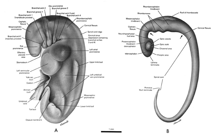


Open PDF version of FIG 5.3, External Features and Nervous System of 5mm Embryo
Figure 5.3 - External Features (A) and Nervous System (B) of 5mm Embryo [PDF version of FIG 5.3]
From Gasser RF, 1975, 49. Atlas of Human Embryos. Copyright © 1975 RF Gasser, PhD. All rights reserved.


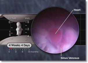
sinus venosus, sinoatrial node, embryo heart, embryonic
Figure 5.4 - Sinus Venosus
The Biology of Prenatal Development DVD
Copyright © 2006 EHD, Inc. All Rights Reserved.
 

By 4 weeks, the heart typically beats between 105 and 121 times per minute.10 The heart’s pacemaker cells, located in the sinoatrial node, develop during this week. These specialized cells originate in the sinus venosus (si’nus ve-no’sus),11 a large vein that collects blood from the entire body as it passes directly into the heart.12 A portion of the sinus venosus including the sinoatrial node becomes part of the right atrium.13 From this location, these pacemaker cells, in cooperation with nerve impulses originating outside the heart, help control a person’s heart rate throughout life.14

The sinus venosus is the final thruway for blood entering the heart.


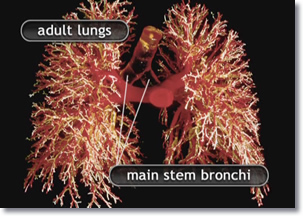
The respiratory system is progressing as 2 primary lung buds form the beginning of the right and left lungs.15 By 4½ weeks, the right and left mainstem bronchi, the major airways to the right and left lungs respectively,16 are well established. They begin dividing into the lobar pattern seen in the adult – 3 lobes on the right and 2 on the left.17

By 5 weeks, repeated branching of the airway system or bronchial (brong’ke-al) tree begins to accelerate. Over the next 12 weeks, this system of airways will undergo most of the 24 divisions present in the adult (although authorities disagree somewhat regarding exactly when these airway divisions are complete.)18 Following birth, these airways connect the air exchange portion of the lungs to the trachea and the outside world.

Baby learning to walk.
At all stages of life, your pacemaker cells help your heart beat faster or slower depending on how active you are.
Frontal view of the adult lungs, main stem bronchi, and bronchial tree.
Figure 5.5 - Adult Lungs - Frontal View
The adult lungs have 3 lobes on the right and 2 lobes on the left. These divisions are already evident just 5 weeks after fertilization.
The Biology of Prenatal Development DVD
Copyright © 2006 EHD, Inc. All Rights Reserved.


Collage of people kick boxing, skating, and playing the flute.
The respiratory system provides our bodies with oxygen needed for all sorts of activities.


By 5 weeks, the embryo’s liver is producing blood cells. This is the first time blood cell formation, or hematopoiesis (he’ma-to-poy-e’sis), begins inside the embryo.19
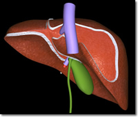
Rear view of the adult liver.
Figure 5.6 - Adult Liver - Rear View
LifeART
Copyright © 2002 Lippincott, Williams & Wilkins.
Play Movie - The Liver and Heart
Movie 5.3 -The Liver and Heart

Development of the stomach, esophagus, pancreas, and the small and large intestines20 is all underway.21

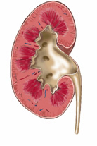
The permanent kidneys appear by 5 weeks.22

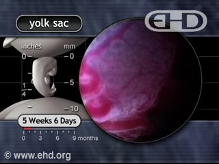
Next to the kidneys, the gonads (go’nads), or reproductive organs, are developing. These will eventually become ovaries in the female and testes in the male. By 5 weeks, early reproductive cells called germ cells begin moving from the yolk sac into the gonads.23 Meanwhile, the yolk sac continues to nourish the embryo until final connections with the placenta form.24
Play Movie - The Yolk Sac
Movie 5.4 - The Yolk Sac
adult human kidney
Figure 5.7 - Adult Human Kidney
The 5-week embryo’s kidneys will eventually look like this adult kidney.
LifeART
Copyright © 2002 Lippincott, Williams & Wilkins.
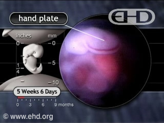
Play Movie - Hand Plates and Cartilage
Movie 5.5 - Hand Plates and Cartilage
Play Movie - Transparent Skin
Movie 5.6 - Transparent Skin
 

The embryo’s endocrine system is also developing. This system of glands regulates the release of hormones throughout a person’s life. The pituitary (pi-tu’i-tar-e) gland forms at the base of the brain during week 5 and begins secreting growth hormone25 and the hormone ACTH which stimulates further growth of the adrenal glands.26


The limb buds continue to grow and by five weeks the embryo develops hand plates.27


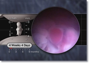
At this point, the embryo’s skin is only one cell thick.28 This makes the skin transparent, allowing us to see internal organs during early development.

Face of sleeping baby.
This baby’s skin hides blood vessels, bones, and organs which were once visible in the womb.
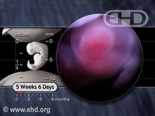
embryo brain, heart, sinus venosus, diaphragm, liver
Figure 5.8 - Embryo Brain, Heart, Sinus Venosus, Liver, and Diaphragm
The transparent skin of this 4 week, 4 day embryo allows easy viewing of the beating heart and liver.
The Biology of Prenatal Development DVD
Copyright © 2006 EHD, Inc. All rights reserved.
back to top
Take a written quiz about this unit. Take a visual quiz about this unit.
4 to 5
Weeks
Footnotes

1 Moore and Persaud, 2003. 440; O'Rahilly and Müller, 1999a. 134; Sadler, 2005. 106.
2 Laffont, 1982. 5.
3 Bartelmez and Dekaban, 1962. 25; Campbell, 2004. 17; O'Rahilly et al., 1984. 249; O'Rahilly and Gardner, 1979. 130; O'Rahilly and Müller, 1999a. 115; van Dongen and Goudie, 1980. 193.
4 Moore, 1980. 938.
5 Guyton and Hall, 2000. 663-677; Noback et al., 1996. 138; O'Rahilly and Müller, 1999b. 41.
6 Moore and Persaud, 2003. 468. Figure 19-2.
7 Gasser, 1975. 50; O'Rahilly and Müller, 1996. 101.
8 O'Rahilly and Müller, 1987. 186.
9 Lecanuet and Schaal, 1996. 12; Moore and Persaud, 2003. 466.
10 van Heeswijk et al., 1990. 153.
11 Moore and Persaud, 2003. 350.
12 O'Rahilly and Müller, 1987. 129; Thompson and Anderson, 1982. 792.
13 Gittenger-de Groot et al., 2000. 20; Moore and Persaud, 2003. 350.
14 Hurst et al., 1990. 22.
15 DiFiore and Wilson, 1994. 221; Fowler et al., 1988. 793.
16 Moore and Persaud, 2003. 245; O'Rahilly and Boyden, 1973. 239; O'Rahilly and Müller, 2001. 291; Sparrow et al., 1999. 550.
17 Moore and Persaud, 2003. 248.
18 DiFiore and Wilson, 1994. 221; Lauria et al., 1995. 466; Moore and Persaud, 2003. 246; Sparrow et al., 1999. 550.
19 Moore and Persaud, 1993. 55.
20 Grand et al., 1976. 793-794, 796, 798.
21 Streeter, 1942. 232, 235.
22 Angtuaco et al., 1999. 13; Lipschutz, 1998. 384; Moore and Persaud, 2003. 288; O'Rahilly and Müller, 1987. 167, 182; O'Rahilly and Müller, 2001. 301; Sadler, 2005. 72.
23 Blackburn, 2003. 7; Moore et al., 2000. 184; O'Rahilly and Müller, 2001. 23; Waters and Trainer, 1996. 16; Witschi, 1948. 70, 77, 79.
24 Campbell et al., 1993. 765; Kurjak et al., 1994. 437.
25 Costa et al., 1993. 625; Matsuzaki et al., 1971. 908.
26 Cunningham FG et al., 2001. 154; Spraycar, 1995. 30.
27 O'Rahilly and Müller, 1987. 175; Streeter, 1948. 139.
28 Cullen et al., 1990. 82-86; Moore and Persaud, 2003. 486; O'Rahilly, 1957. 459; O'Rahilly and Müller, 2001. 165.