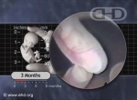
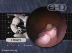
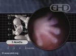
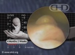
Prenatal Summary
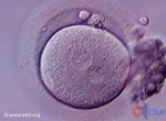
Pregnancy begins at conception with the union of a man’s sperm and a woman’s egg to form a single-cell embryo.1This brand new embryo contains the original copy of a new individual’s complete genetic code. Gender,2 eye color, and other traits are determined at conception, also known as fertilization.
Distributed byMost significant developmental milestones occur long before birth during the first eight weeks following conception when most body parts and all body systems appear and begin to function.3
The main divisions of the body, such as the head, chest, abdomen and pelvis, and arms and legs are established by about four weeks after conception.4 Eight weeks after conception, except for the small size, the developing human’s overall appearance and many internal structures closely resemble the newborn.5
Pregnancy is not just a time for growing all the parts of the body. It is also a time of preparation for survival after birth.6 Many common daily activities seen in children and adults begin in the womb—starting more than 30 weeks before birth. These activities include hiccups, touching the face, breathing motions, urination, right- or left-handedness, thumb sucking, swallowing, yawning, jaw movement, reflexes, REM sleep, hearing, taste, sensation, and so on.
Full-term pregnancy typically lasts 38 weeks from conception or 40 weeks from the first day of a woman’s last normal menstrual period.7
Unless otherwise noted, all prenatal ages on this web page are referenced from the start of the last normal menstrual period. This age is two weeks greater than the age from conception, also referred to as fertilization. (Please note that on the remainder of ehd.org, prenatal ages are referenced from the time of conception.)
The First Two Weeks
Shortly after a woman’s period begins, her body begins preparing for the possibility of pregnancy.
Approximately 2 weeks into her cycle, a woman releases an egg from one of her ovaries into her adjacent fallopian tube. Conception is now possible for the next 24 hours or so8 and signifies the beginning of pregnancy.9
The single-cell embryo has a diameter of approximately 4 thousandths of an inch.10
2 to 4 Weeks
The cells of the embryo repeatedly divide as the embryo moves through the Fallopian tube into the woman’s uterus or womb. Implantation, the process whereby the embryo embeds itself into the wall of the womb, begins by the end of the third week and is completed during the fourth week of pregnancy.11
The 4-week embryo is less than 1/100th of an inch long.
4 to 6 Weeks
By 5 weeks, development of the brain, spinal cord,12 and heart13 is well underway.
The heart begins beating at 5 weeks and one day14 and is visible by ultrasound almost immediately.15
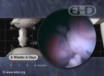
By 6 weeks, the heart is pumping the embryo’s own blood to his or her brain and body.16 All four chambers of the heart are present17 and more than 1 million heartbeats have occurred.18 The head, as well as the chest and abdominal cavities have formed19 and the beginnings of the arms and legs are easily seen.20
The 6-week embryo measures less than ¼ of an inch long from head to rump.
6 to 8 Weeks
Rapid brain development continues with the appearance of the cerebral hemispheres at 6½ weeks.21
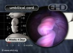
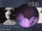
The embryo reflexively turns away in response to light touch on the face at 7½ weeks.22
Fingers are beginning to form on the hand.23
By 8 weeks the developing human measures about a ½ inch from head to rump.
8 to 10 Weeks
Brainwaves have been measured and recorded before 8½ weeks.24
Also by 8½ weeks, the bones of the jaw and collar bone begin to harden.25
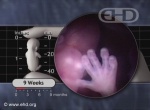
By 9 weeks the hands move, the neck turns,26 and hiccups begin.27 Girls now have ovaries28 and boys have testes.29 The embryo’s heart rate peaks at about 170 beats per minute30 and will gradually slow down until birth.
Electrical recordings of the heart at 9½ weeks are very similar to the EKG tracing of a newborn.31 The heart is nearly fully formed.32
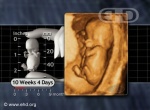
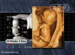
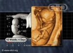
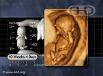
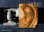
By 10 weeks kidneys begin to produce and release urine,33 and intermittent breathing motions begin.34 All fingers and toes are free and fully formed,35 and several hundred muscles are present.36 The hands and feet move frequently and most embryos show the first signs of right- or left-handedness.37
Experts estimate the 10-week embryo possesses approximately 90% of the 4,500 body parts found in adults.38 This means that approximately 4,000 permanent body parts are present just eight weeks after conception.
Incredibly, this highly complex 10-week embryo weighs about 1/10th of an ounce and measures slightly less than 1¼ inches from head to rump.
10 to 12 Weeks
After 10 weeks, the developing human is called a fetus, which means “little one” or “unborn offspring.”39
The eyelids are temporarily fused together by 10½ weeks.40
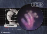
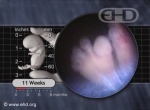
By 11 weeks the head moves forward and back, the jaw actively opens and closes, and the fetus periodically sighs41 and stretches.42 The face, palms of the hands, and soles of the feet are sensitive to light touch.43 Thumb sucking44 and swallowing amniotic fluid begin.45 Girls’ ovaries now contain reproductive cells46 which will give rise to eggs later in life. Also in girls, the uterus is now present.47
Yawning begins at 11½ weeks.48
The number of heartbeats now exceeds 10 million.
Fingerprints start forming at 12 weeks49 while fingernails and toenails begin to grow.50
The bones are hardening in many locations.51
The 12-week fetus weighs less than 1 ounce and measures about 3 inches from head to heel.
12 to 14 Weeks
By 13 weeks the lips and nose are fully formed52 and the fetus can make complex facial expressions.53
By 14 weeks taste buds are present all over the mouth and tongue.54
The fetus now produces a wide variety of hormones.55
Arms reach final proportion to body size.56
The 14-week fetus weighs about 2 ounces and measures slightly less than 5 inches from head to heel.
14 to 16 Weeks
By 15 weeks the entire fetus (except for parts of the scalp) responds to light touch.57 Tooth development is underway.58
Gender differences emerge at 16 weeks when girl fetuses move their jaws more often than boys.59
A pregnant woman may begin to feel fetal movement at this time.60
The 16-week fetus weighs about 4 ounces and measures slightly less than 7 inches from head to heel.
16 to 18 Weeks
Production of a variety of digestive enzymes is well underway.61
Around 17 weeks blood cell formation moves to its permanent location inside the bone marrow62 and the fetus begins storing energy in the form of body fat.63
By 18 weeks formation of the breathing passages, called the bronchial tree, is complete.64 The fetus releases stress hormones in response to being poked with a needle.65
The 18-week fetus weighs around 6 ounces and measures about 8 inches from head to heel.
18 to 20 Weeks
By 19 weeks, more than 20 million heartbeats have occurred.
By 20 weeks the larynx or voice box begins moving in a way similar to the movement seen during crying after birth.66 The skin has developed sweat glands67 and is covered by a greasy white substance called “vernix,”68 which provides protection from the amniotic fluid.69
The 20-week fetus weighs about 9 ounces and measures about 10 inches from head to heel.
20 to 22 Weeks
At 21 weeks breathing patterns, body movements, and heart rate begin to follow daily cycles called circadian rhythms.70
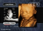
By 22 weeks the sense of hearing begins to function and the fetus starts responding to various sounds.71 The cochlea, the organ of hearing, reaches adult size.72 All skin layers and structures are complete.73
With specialized medical care some fetuses can survive outside the womb by 22 weeks with survival rates reported as high as 40%74 in some medical centers.
The 22-week fetus weighs slightly less than 1 pound and measures about 11 inches from head to heel.
22 to 24 Weeks
Between 20 and 23 weeks rapid eye movements begin. These eye movements are similar to those seen when children and adults have dreams.75
By 24 weeks more than 30 million heartbeats have occurred.
The 24-week fetus weighs about 1¼ pounds and measures about 12 inches from head to heel.
24 to 26 Weeks
By 25 weeks, breathing motions may occur up to 44 times per minute.76
By 26 weeks sudden, loud noises may trigger a blink-startle response,77 which may increase movement, heart rate, and swallowing.78
The lungs produce a substance necessary for breathing after birth.79
The 26-week fetus weighs almost 2 pounds and measures about 14 inches from head to heel.
26 to 28 Weeks
By 27 weeks the thigh bones and the foot bones are each about two inches long (about 5 cm).80
By 28 weeks the sense of smell is functioning81 and eyes produce tears.82
The 28-week fetus weighs more than 2½ pounds and measures about 15 inches from head to heel.
28 to 30 Weeks
By 29 weeks, pupils of the eyes react to light.83
The 30-week fetus weighs about 3¼ pounds and measures about 16 inches from head to heel.
30 to 32 Weeks
By 31 weeks more than 40 million heartbeats have occurred. Wrinkles in the skin are disappearing as more and more fat deposits are formed.84
By 32 weeks breathing movements occur up to 40 percent of the time.85
The 32-week fetus weighs about 4 pounds and measures about 17 inches from head to heel.
32 to 34 Weeks
By 34 weeks true alveoli, or "air pocket" cells, begin developing in the lungs.86
The 34-week fetus weighs about 5 pounds and measures about 18 inches from head to heel.
34 to 36 Weeks
The 36-week fetus weighs about 5¾ pounds and measures about 18½ inches from head to heel.
36 to 38 Weeks
By 37 weeks the fetus has a firm hand grip87 and the heart has beat more than 50 million times.
The 38-week fetus weighs about 6¾ pounds and measures about 19 inches from head to heel.
38 to 40 Weeks
At term, the umbilical cord is typically 20 to 24 inches long.88
Labor is initiated by the fetus,89 ideally around 40 weeks, leading to childbirth.
At full-term birth, newborn babies typically weigh between 6 and 9 pounds and measure between 18 and 21 inches from head to heel.90
Free licensed content for government health and education departments
The Endowment for Human Development, Inc. (EHD) hereby grants state and local governments a non-exclusive limited license to incorporate any or all of the prenatal development facts presented on this “Prenatal Summary” web page (English and Spanish) into government web pages and printed materials at no charge. Government entities using EHD material on the Internet are required to acknowledge EHD in the credits and provide at least one live link to this Prenatal Summary web page (https://www.ehd.org/prenatal-summary.php). Similarly, governments using EHD development facts in printed materials should list EHD as a source in the credits along with the web address of this page.
Free embeddable video clips are also available for government and school web pages in English and Spanish. Links to EHD’s Image Gallery and Movie Theater are welcome.
Additionally, EHD will review your prenatal development teaching materials and provide feedback upon request at no charge (subject to availability). Please submit questions to .
Footnotes
| 1 | Carlson, 2009. 3. |
| 2 | Carlson, 2009. 35; Gasser, 1975. 1; Guyton and Hall, 2000. 944-945. |
| 3 | Moore and Persaud, 2003. 3. |
| 4 | Carlson, 2009. 123; O'Rahilly and Müller, 1987. Figure 13-2, 142. |
| 5 | O'Rahilly and Gardner, 1975. 15; O'Rahilly and Müller, 1999a. 228. |
| 6 | Vindla and James, 1995. 598; Carlson, 2009. 490. "Fetal breathing movements are essential for postnatal survival. One function of fetal breathing is to condition the respiratory muscles so they can perform postnatal contractions." |
| 7 | Carlson, 2009. 23; Cunningham FG et al., 2001. 226; O'Rahilly and Müller, 2001. 92. |
| 8 | Sadler, 2005. 6; Cunningham FG et al., 2001. 86. |
| 9 | Carlson, 2009. 3. |
| 10 | Unless otherwise noted, all measurements of the weight and overall length of the embryo and fetus in this summary are derived from this source: O'Rahilly and Müller, 2001. Table A-1, 490. |
| 11 | Implantation begins with attachment of the embryo about 6 days after fertilization. [Attachment of the embryo to the inner wall of the uterus is a transient event and is the hallmark of Carnegie Stage 4.] O'Rahilly and Müller, 2001. 40; Adams, 1960. 13-14. |
| 12 | Bartelmez, 1923. 236; Müller and O'Rahilly, 1983. 419-420, 429; O'Rahilly and Gardner, 1979. 123, 129; O'Rahilly and Müller, 1984. 422; O'Rahilly and Müller, 1987. 90; O'Rahilly and Müller, 1999a. 47, 52. |
| 13 | O'Rahilly and Müller, 1987. 86; Gilmour, 1941. 28. |
| 14 | Campbell, 2004. 14; Carlson, 2004. 430; de Vries and Saunders, 1962. 96; Gardner and O'Rahilly, 1976. 583; Gilbert-Barness and Debich-Spicer, 1997. 650; Gittenger-de Groot et al., 2000. 17; van Heeswijk et al., 1990. 151; Kurjak, Chervenak, 1994. 439; Navaratnam, 1991. 147-148; O'Rahilly and Müller, 1987. 99; Wisser and Dirschedl, 1994. 108. |
| 15 | Kurjak, Chervenak, 1994. 439. This source cites Neiman HL, 1990 as having documented cardiac activity at 36 postmenstrual days or 22 postfertilization days; Wisser and Dirschedl, 1994. 108. They reported using transvaginal ultrasound to visualize the embryonic heartbeat 23 days postfertilization in two embryos fertilized in vitro "with exactly known age" and "in embryos from 2 mm of greatest length onwards." |
| 16 | O'Rahilly and Müller, 1987. 143. |
| 17 | Gasser and Cork, 2001-2011. Stage 13, sections 112-113, 131-132. |
| 18 | EHD, 2005; EHD, 2010. All estimates of the number of heartbeats at various times during pregnancy are derived from these sources and will not be repeated in this article. |
| 19 | Gasser and Cork, 2001-2011. Carnegie Stage 13. |
| 20 | Gasser, 1975. 49, 59; O'Rahilly and Gardner, 1975. 11; O'Rahilly and Müller, 1985. 148, 151; O'Rahilly and Müller, 1987. 143; Streeter, 1945. 30; Uhthoff, 1990. 7, 141. |
| 21 | Bartelmez and Dekaban, 1962. 25; Campbell, 2004. 17; O'Rahilly and Gardner, 1979. 130; O'Rahilly et al., 1984. 249; O'Rahilly and Müller, 1999a. 115; van Dongen and Goudie, 1980. 193. |
| 22 | Goodlin, 1979. D-128; Humphrey, 1964. 99. |
| 23 | Moore and Persaud, 2008. Table 5-1, 80. Precursors to individual fingers and metacarpal bones (called digital rays) are first seen; Gasser and Cork, 2001-2011. Carnegie Stage 17, sections 541, 547, and 553. |
| 24 | Borkowski and Bernstine, 1955. 363. (cited by Bernstine, 1961, 63 & 66; O'Rahilly and Müller, 1999a, 195; and van Dongen and Goudie, 1980, 193.); Hamlin, 1964. 287. For a summary of in utero fetal encephalography (measuring brainwaves) in the near-term fetus using abdominal and vaginal electrodes see Bernstine et al., 1955. |
| 25 | Carlson, 2004. 189; O'Rahilly and Gardner, 1972. 293; O'Rahilly and Gardner, 1975. 19; O'Rahilly and Müller, 2001. 385; Sperber, 1989. 122, 147. |
| 26 | EHD, 2006. See the embryo in action. |
| 27 | de Vries et al., 1982. 305, 311; Visser et al., 1992. Figure 1, 176. |
| 28 | O'Rahilly and Müller, 2001. 320. |
| 29 | O'Rahilly and Müller, 1987. 254. |
| 30 | van Heeswijk et al., 1990. 153; Leeuwen et al., 1999. 265; van Lith et al., 1992. 741. Various authors agree the heart rate peaks at 7 weeks. Reported heart rates vary however. Van Heeswijk et al. report a peak heart rate of 167 ± 8 beats per minute (bpm) while Leeuwen et al. report a peak rate of 175 bpm. Van Lith et al. report the median fetal heart rate peaks at 177 bpm at 7 weeks. |
| 31 | Straus et al., 1961. 446. (cited by Gardner and O'Rahilly, 1976, 571.): "…an electrocardiogram with the classical P, QRS, and T configuration has been obtained from a 23mm human embryo (Straus, Walker, and Cohen, 1961)." |
| 32 | Cooper and O'Rahilly, 1971. 292; James, 1970. 214; Jordaan, 1979. 214; Streeter, 1948. 192; Vernall, 1962. 23. "The four chambers of the heart and the associated major vessels are externally apparent in a close approximation to their adult positions." |
| 33 | O'Rahilly and Müller, 2001. 304; Windle, 1940. 118. (Windle reports urine formation begins at nine weeks postfertilization or 11 postmenstrual weeks.) |
| 34 | Connors et al., 1989. 932; de Vries et al., 1982. 311; McCray, 1993. 579; Visser et al., 1992. 177. |
| 35 | O'Rahilly and Müller, 1987. Figure 23-1, 260; Gasser and Cork, 2001-2011. Stage 23. |
| 36 | Gasser and Cork, 2001-2011. Stages 19 through 23. |
| 37 | Hepper et al., 1998. 531; McCartney and Hepper, 1999. 86. |
| 38 | O'Rahilly and Müller, 2001. 87. "It has been estimated that more than 90% of the more than 4500 named structures of the adult body become apparent during the embryonic period (O'Rahilly)." |
| 39 | Moore and Persaud, 2008. 2. |
| 40 | Andersen et al., 1965. 648-649; O'Rahilly and Müller, 2001. 465; Pearson, 1980. 39-41. |
| 41 | de Vries et al., 1982. 305-307. |
| 42 | de Vries et al., 1982. 311. |
| 43 | Humphrey, 1964. 96; Humphrey, 1970. 16-17. (cited by Reinis and Goldman, 1980, 232); Humphrey and Hooker, 1959. 77-78. |
| 44 | Liley, 1972. 103. |
| 45 | Campbell, 2004. 24; de Vries et al., 1982. 311; Petrikovsky et al., 1995. 605. |
| 46 | O'Rahilly and Müller, 2001. 25, 322. |
| 47 | O'Rahilly and Müller, 2001. 327; O'Rahilly, 1977b. 53; O'Rahilly, 1977a. 128. |
| 48 | de Vries et al., 1982. 309. |
| 49 | Babler, 1991. 95; Penrose and Ohara, 1973. 201; For an overview of ridge formation in the skin of the hands see: Cummins, 1929. 103-126. |
| 50 | O'Rahilly and Müller, 2001. 170-171. |
| 51 | Cunningham FG et al., 2001. 133. |
| 52 | Timor-Tritsch et al., 1990. 291. |
| 53 | Valman and Pearson, 1980. 234. |
| 54 | Lecanuet and Schaal, 1996. 3; Miller, 1982. 169; Mistretta and Bradley, 1975. 80; Carlson, 2009. 493. |
| 55 | Carlson, 2009. 494. |
| 56 | Moore and Persaud, 2003. 105. |
| 57 | Lecanuet and Schaal, 1996. 2; Reinis and Goldman, 1980. 232. |
| 58 | Pringle, 1988. 178. |
| 59 | Hepper et al., 1997. 1820. |
| 60 | Sorokin and Dierker, 1982. 720; Leader, 1995. 595. |
| 61 | Carlson, 2009. 493; Grand et al., 1976. 797. |
| 62 | Pringle, 1988. 179. |
| 63 | Poissonnet et al., 1983. 7; Poissonnet et al., 1984. 3. In a study of 488 fetuses, Poissonnet's group found that adipose tissue (fat) appears in the face starting in the 14th week postfertilization. By 15 weeks, fat appears in the abdominal wall, back, kidneys, and shoulders. By 16 weeks, fat is also present throughout the upper and lower limbs. |
| 64 | DiFiore and Wilson, 1994. 221-222. [There is some disagreement among experts regarding when the bronchial tree is complete. Some say completion occurs as early as 16 weeks postfertilization while others say it occurs after birth.]; Pringle, 1988. 178. |
| 65 | Giannakoulopoulos et al., 1999. 494, 498-499; Glover and Fisk, 1999. 883; Smith et al., 2000. 161; Also of interest, cortisol levels also rise after invasive procedures following 21 weeks postfertilization - see: Giannakoulopoulos et al., 1994. 80. |
| 66 | Lopez Ramon y Cajal, 1996. 65. |
| 67 | Pringle, 1988. 180. |
| 68 | Moore et al., 2000. 52. |
| 69 | Campbell, 2004. 48; Moore and Persaud, 2003. 107; O'Rahilly and Müller, 2001. 168. |
| 70 | de Vries et al., 1987. 333; Goodlin and Lowe, 1974. 349; Okai et al., 1992. 391, 396; Romanini and Rizzo, 1995. 121; For a description of the circadian system, see: Rosenwasser, 2001. 127; Vitaterna et al., 2001. 92. Glossary: "Circadian: A term derived from the Latin phrase "circa diem," meaning "about a day;" refers to biological variations or rhythms with a cycle of approximately 24 hours." |
| 71 | Glover and Fisk, 1999. 882; Hepper and Shahidullah, 1994. F81; Querleu et al., 1989. 410; Sorokin and Dierker, 1982. 725, 730; Valman and Pearson, 1980. 233-234. |
| 72 | Lecanuet and Schaal, 1996. 5-6; Querleu et al., 1989. 410. |
| 73 | Pringle, 1988. 180. |
| 74 | Zayek et al., 2011. Table 1, 128. |
| 75 | Okai et al., 1992. 392. |
| 76 | Natale et al., 1988. 317. |
| 77 | Birnholz and Benacerraf, 1983. 517; Campbell, 2002. 3. Professor Stuart Campbell correctly points out that the eyes of the fetus are closed most of the time and a true blink requires the eyes to be open. Perhaps the "blink-startle" response would be more accurately termed "squint-startle."; Drife, 1985. 778. |
| 78 | Visser et al., 1989. 285; Petrikovsky et al., 1993. 548-549. |
| 79 | Moore et al., 2000. 154; Carlson, 2009. 490. |
| 80 | Cunningham FG et al., 2001. Figure 41-4, 1116-1117; Unless otherwise noted, all measurements of the weight and overall length of the embryo and fetus in this summary are derived from this source: O'Rahilly and Müller, 2001. Table A-1, 490. |
| 81 | Lecanuet and Schaal, 1996. 3. |
| 82 | Isenberg et al., 1998. 773-774. |
| 83 | Robinson and Tizard, 1966. 52. |
| 84 | England, 1983. 28. |
| 85 | Connors et al., 1989. 932; de Vries et al., 1985. 117; Patrick et al., 1980. 26, 28; Visser et al., 1992. 178. |
| 86 | Lauria et al., 1995. 467. |
| 87 | Moore and Persaud, 2003. 108. |
| 88 | Carlson, 2009. 139. |
| 89 | Liley, 1972. 100. |
| 90 | Clinical Growth Charts, 2001. 1. |


