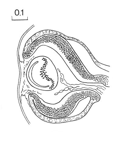
Cross-section of the developing eye. At this stage the internal neuroblastic layer of retina is being formed by migration. At this time the migration is limited to a restricted field, approximately at the site of the macula.
Also during this stage the lens disc becomes thickened with the histogenesis of lens fibers. The lens disc begins to fill the lens vesicle, reducing it to a crescent-shaped cleft.
O'Rahilly and Müller, 1987 Fig. 17-14
Keywords: crescent-shaped cleft, eye, lens disc, lens fiber(s), lens vesicle, macula, neuroblastic layer
Source: The Virtual Human Embryo.
