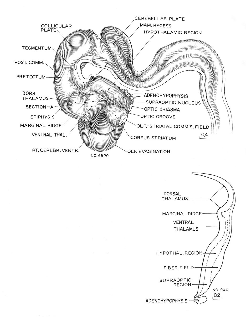
Reconstruction of the right half of brain of an embryo No. 6520. The marginal ridge, projecting into the lumen and separating the dorsal thalamus from the ventral thalamic region lying rostral to it, is now plainly seen. The contours of the wall are seen in section A. This drawing was made from No. 940 of the same stage, and it corresponds to a transverse section along the dotted line shown in the sketch of the reconstruction, passing caudal to the epiphysis, with its lower end transecting the hypophysis. It will be noted that above the marginal ridge the wall is behind in development, and ventral to it the wall is precocious. The ventral thalamic region has a nuclear area comparable to a mantle zone. In the hypothalmic region there is a large fiber field which appears to unite later with the peduncular system. The neurohypophysis forms a deep ventral pocket with characteristic foldings of its caudal wall. The adenohypophysis is flattened dorsoventrally, but spreads widely, enclosing the infundibulum on each side with its two wings: it is therefore larger than is shown in a median section. Finally, it is to be noted that the olfactory evagination is just making its appearance.
O'Rahilly and Müller, 1987 Fig.17-12
Keywords: adenohypophysis, cerebellar plate, collicular plate, corpus striatum, dorsal thalamus, epiphysis, fiber field, hypophysis, hypothalamus, infundibulum, mamillary recess, marginal ridge, olfactory evagination, olfactory-striatal commissural field, optic chiasma (chiasmatic plate), optic groove, posterior communicating artery, pretectum, right cerebral ventricle, right half of the brain, supra-optic nucleus, supra-optic region, tegmentum, ventral thalamus
Source: The Virtual Human Embryo.
